Every year in Ireland, an average of 65 children and teenagers, are diagnosed with a tumour of the brain or spinal cord. Brain tumours are the most common tumours seen in children, and account for 27% of childhood cancer every year, alongside leukaemia (also 27%).
A diagnosis like this is of course, a very stressful time for the entire family.
This information is written for you, the parent or guardian, please also refer to our new ‘Heads Up’ guide below:
- The brain and spinal cord, which is called the central nervous system (CNS)
- The types of tumours that occur in this area
- The different ways that these tumours are treated
We wish to thank the doctors and nurses at Beaumont Hospital, Crumlin Children’s Hospital and Temple Street Hospital for their work on this information.
There are many different types of tumours and each patient is unique. This means that even the same type of tumour can affect different children in different ways, and the treatments may not be identical.
The doctors and nursing staff looking after your child will give you individual information about your child’s condition and are best placed to answer any specific questions or discuss any concerns you may have.

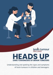
Heads Up | Early Diagnosis of Brain Tumours
This year, as part of National Brain Tumour Awareness Week 2025, Brain Tumour Ireland have developed a new guide to help spot the signs and symptoms of brain tumours in children and teenagers.
Thanks to Children’s Hospital Ireland (CHI) and The Brain Tumour Charity UK for their contribution.
You can view/ download this booklet by clicking here.

Symptoms are different in children
- Persistent vomiting/feelings of nausea (over a two week period)
- Recurring headache (over a four week period, particularly on waking)
- Abnormal eye movements
- Fits or seizures
- Behaviour change
- Abnormal balance/walking/co-ordination
- Blurred/double vision
- Abnormal head position (such as a head tilt)
- Delayed or arrested puberty (puberty that doesn‘t start or starts, but doesn’t progress as expected)
Diagnosis & symptoms
Our bodies are made up of billions of tiny building blocks called cells. Each cell has a specific role to play, and contains our own DNA. In normal day to day life, our cells are constantly “turning over”, which means that they are doing their job, dying off, and being reproduced by new versions of themselves. This process sometimes results in cells with “mistakes” in them. This is common, and happens in all of us. Our immune system usually destroys cells with mistakes in them. However, for a variety of complex reasons, sometimes a cell with a mistake in it can escape from the immune system. This can result in the cell dividing and multiplying in an uncontrolled way and forming a tumour. If this happens in the brain it is called a brain tumour; if it occurs in the spine it is a spinal tumour. The name of each specific tumour, depends on the exact type of cell that grew out of control initially.
People use many different words for tumours, which can be confusing. A tumour is an abnormal group of cells forming a lump or mass. Often people may refer to a tumour as a lump or a mass. A tumour may be benign (not cancerous) or malignant (cancerous). The only way of knowing with certainty, if a tumour is benign or malignant, is by removing a piece of it (a biopsy), or all of it (a resection) and examining it in the laboratory. This is a complex process that involves many different steps. This is why it can take 7-10 days (and sometimes longer) to know an exact diagnosis.
Brain tumours that occur in infants and children are very different from adult brain tumours, both in terms of the type of cells involved and their responsiveness to treatment. Brain tumours in children usually originate there (primary brain tumours); as opposed to adults who often have brain tumours which have spread from other parts of the body (secondary brain tumours). Tumours in the brain or spinal cord can sometimes spread (metastasise), but this spread is almost always confined to within the Central Nervous System (brain and spinal cord). The cause of childhood brain tumours is unknown in the majority of cases. There are a small number of children who have a genetic predisposition for developing tumours e.g. neurofibromatosis, tuberous sclerosis, and Li-Fraumeni syndrome. If your oncologist believes that your child potentially has one of these conditions, they will discuss this with you; and together, you will make the decision, to consider testing for these conditions where appropriate. Children, teenagers or young adults who have been exposed to radiotherapy in the past have an increased risk of developing tumours in the previously irradiated site. Some radiation induced tumours are benign, and some may be malignant.
The first stage of diagnosis starts when your child is seen by your GP or local hospital. This is usually because your child has had a specific symptom, or some vague ones, that caused you concern and need to be investigated. Brain tumours are generally first discovered on a brain scan (CT or MRI). Once a brain tumour is suspected, your child is referred toa specialist team of doctors and nurses.
Neurosurgery is a very important component of the care of children or teenagers with brain or spinal cord tumours. The neurosurgeon will often be the first specialist from the specialist team to meet your family. He or she will discuss with you the symptoms you have noticed in your child and explain what was seen on the scan. It is likely that further tests/investigations/procedures are required, so that information can be gathered in a safe way; in order to explain an accurate diagnosis to you and your family.
The care of children, teenagers and young adults with tumours of the brain or spinal cord, involves a large team of specialists. We work together very closely in order to:
• Safely make an accurate diagnosis
• Create and deliver a tailored treatment plan
• Provide follow-up care after therapy
• Support the family throughout their journey
Members of The National Children’s Cancer Service (NCCS), currently based in Crumlin, together with The National Department of Neuro-surgery (currently based in Temple Street) work together to form the paediatric neuro-oncology multi-disciplinary team. This expert team includes neuro-surgeons, paediatric oncologists, radiation oncologists, radiologists, neuro-pathologists, and a dedicated nursing team. We have fostered close links with dedicated social workers, psychologists, physiotherapy, occupational therapy, speech and language therapy, dietetics, school, and complementary therapists, who provide a fundamental aspect of our service. Many other teams are involved, as their expertise is required.
Complete medical history: A doctor will sit down with you and your child and ask detailed questions about the symptoms that you have noticed recently. It is usual to also ask questions about the child’s previous medical history, birth history, family history, and developmental history etc.
Clinical Examination: Your child’s doctor will examine them at the bedside. This does not usually hurt, but some children dislike it, particularly if they are feeling unwell. Your child will have a detailed examination including a neurological examination .This involves testing reflexes, muscle strength, eye and mouth movement, coordination, and alertness.
A Computerised Tomography scan (CT or CAT scan): A CT scan is a detailed scan which looks at the whole brain. It can be used to look for tumours, swelling, bleeding and fluid problems. The CT scanner is shaped like a large Polo mint and has a table which sits inside the ring shape. Your child will need to lie still on this table. A special dye will usually be injected into their vein to give a clearer picture of their brain. A CT scan usually lasts about 15–20 minutes. For this reason younger children may need sedation or a short anaesthetic. A CT scan is associated with a small dose of radiation.
Magnetic Resonance Imaging (MRI): An MRI scan is a type of scan that uses a very strong magnet and radio-waves to give more information about the position of the tumour. Your child will need to lie still on a table in a special X-ray tunnel for 30 minutes or longer. You should prepare your child in advance for this scan as the machine is very noisy (hammering sound) and they may find it difficult to lie still in the tunnel. However, usually the children can listen to music or look at DVD to distract them until the scan is completed. Very young children are given a general anaesthetic for this type of scan. Older children who are prepared in advance tend to cope very well, but parents or guardians can also remain in the room with them during the scan. Because the scan uses magnets, all metal objects (jewellery, clips or pins) will have to be removed. If your child wears dental braces, this may make the MRI image less clear. You may be advised to have them temporarily removed for the scan. You will also need to tell staff if your child has had any metal devices implanted in their body in the 6 weeks before the scan, e.g. a metal pin for a broken leg, as the device may not yet be secure and staff may need to take special safety measures.
Ophthalmology (eye) and Audiology (hearing) examination:
Various eye and hearing tests may be performed as part of the assessment for certain tumours.
Lumbar puncture/spinal tap
A lumbar puncture involves taking a sample of fluid surrounding the spinal cord for examination. When a lumbar puncture is performed, a fine needle is carefully inserted into the lower back to take a sample of fluid. This is usually done under sedation or general anaesthetic to minimise discomfort. This test is to show if the tumour cells have spread/metastasised to the CSF. The CSF may also be analysed for chemicals called tumour markers, which certain types of brain tumours may leak into the CSF.
Bloods tests
Certain tumours called germ cell tumours release chemicals that can be found in the blood and/or CSF. If a germ cell tumour is suspected, a blood test can be taken to look for these chemicals. It is sometimes possible to diagnose the type of tumour in this way without the need for a tumour biopsy.
EEG
An EEG or electroencephalogram is a test that measures the electrical activity in the brain. The test may be done for your child if their tumour is causing seizures or fits (A fit is another word for a seizure). It involves placing small electrodes on their scalp, which transmit and record electrical brain activity onto a graph for the doctors to read. This test does not hurt, although children may complain about the sticky gel in their hair. The gel is used to help keep the electrodes in place.
Endocrine (hormone) Tests
The pituitary gland is the master hormone gland in the body, and responsible for the release of regulating hormones. Certain tumours and their treatments can sometimes interfere with the normal hormone changes that happen as your child grows. Depending on the type and location of your child’s tumour, and the type of treatment that they’ve had, they may need their hormonal system monitored closely during childhood and adolescence. This will be done at outpatient follow up clinics and usually involves physical examination and blood tests.
The vast majority of brain and spinal cord tumours require a biopsy or removal (resection) of the tumour, in order to make a diagnosis (give the tumour a name).
Biopsy
In some situations when it is not possible, or perhaps not immediately necessary to remove the tumour, doctors may need to remove a small piece of the tumour to find out exactly what it is. Your child will be given an anaesthetic for the procedure and it will be done in the operating theatre.
Tumour Resection (removal)
Sometimes, it’s necessary to remove the tumour. This may require more than one surgery. The neuro-surgical team will explain to you what type of surgery is required, the potential side-effects for your child, and the likely recovery time.
The tumour sample is then sent to the neuropathology team, who start the process of testing, to find out what type of tumour it is. A neuropathologist is a specialist doctor who examines tumour cells in the laboratory. He or she will make a diagnosis based on the sample. On average, a result can take 7 to 10 days. Sometimes the results may take longer to process depending on the complexity of the analysis. Specialist molecular (DNA profiling) results often take weeks to months to obtain. Once a diagnosis has been made all members of the specialist team work together to ensure that your child gets the most appropriate treatment in a timely way. In general, children need to recover from their surgery prior to starting other therapies.
Other Surgical interventions:
If your child’s tumour is blocking the cerebrospinal fluid (CSF) channels in their brain; this can cause an increase in pressure, which is called hydrocephalus. Hydrocephalus may result in headache, vomiting, sleepiness and abnormal eye movements. There are a variety of ways to treat this and the neuro-surgical team will recommend the most appropriate plan for your child. Hydrocephalus may be treated by tumour removal, insertion of specialised devices called shunts; or by creating a new channel called an endoscopic third ventriculostomy (ETV).
The first signs of a brain tumour are variable, and depend on many factors; including, the precise location of the tumour, the structures the tumour is putting pressure on, and the age of the child. It is very common for the initial signs and symptoms to be mistaken for more common childhood illnesses e.g. a tummy bug or migraine. We sometimes meet children or teenagers who have brain tumours and are completely well, but were having a scan of their head for another reason; this is called an incidental finding.
The HEADSMART initiative was developed in the UK, and aimed to increase population awareness regarding brain tumours in children. This symptom card was designed to help you know and spot the signs and symptoms of brain tumours in children and teenagers.
Treatment
Your child will have their diagnosis and treatment plan recommended by the neuro-oncology multi-disciplinary team, which includes experts from all relevant specialties. The paediatric oncologist will discuss this treatment plan in detail with you and answer all of your questions. Each child’s treatment plan is unique to them, but will be based on “standard of care”. This means that the treatment plan which is recommended will be based on available scientific evidence and expertise, and is the “best way we know how” to treat a particular tumour type. Sometimes there is an opportunity to enrol on a clinical trial, which will be discussed with you.
Selecting treatments to treat children with brain and spinal cord tumours is always a very careful balance; that is, we aim to pick treatment which maximises our chance of cure but minimises the chances of long term effects from the tumour and the treatments. These decisions are often straightforward, but sometimes require complex discussion. Please be reassured that your medical team is committed to answering all of your questions, as we know this is a very worrying time for your family.
See under diagnosis section.
Brain or spinal cord tumours that are not causing significant symptoms for your child, and are believed to be slow growing are often “observed” closely. This involves regular outpatient visits and MRI scans to ensure that the tumour is not showing signs of growth. There are many children who are looked after in this way, and never require intervention. If the tumour starts to grow or cause symptoms for your child, further discussion is had regarding next steps required.
Chemotherapy is a term used to describe many different types of medicines used to treat tumours (both cancerous and non-cancerous). Chemotherapy is sometimes given as a tablet or liquid in the mouth, into a vein (intravenously) or into the cerebrospinal fluid (intrathecal). Chemotherapy affects cells that are rapidly dividing, like tumour cells. The aim of giving chemotherapy is that it stops or slows down tumour growth. If a tumour has been removed completely, we often still give chemotherapy, with the aim of “mopping up” tiny tumour cells that are left over. There are many different types of chemotherapy drugs which may be used alone or in combination with others, depending on the type of tumour being treated.
Throughout the world paediatric oncologists work together to develop successful treatment plans, which are also called protocols. Children are treated on a specific protocol depending on their tumour type and age. The paediatric oncologist will explain to you in detail which protocol is suitable for your child and any possible side-effects associated with that treatment. Chemotherapy protocols are usually given over many months, with children attending the Oncology Unit at regular intervals, either as a day-patient or in-patient.
Chemotherapy is usually given by drip into a vein (intravenously) or in tablet or liquid form. For some tumours, chemotherapy is injected directly into the cerebrospinal fluid surrounding the brain and spinal cord. This is called intrathecal chemotherapy. A device called an Ommaya reservoir is sometimes used to give intrathecal chemotherapy.
Radiotherapy is a treatment that uses high-energy radiation to treat tumours. Treatment is like having an x-ray taken and is completely painless. It is usually directed very accurately at the tumour to destroy the cancer cells. Radiotherapy is given as a course of daily treatments, over a period of weeks. It is usually used in combination with surgery and/or chemotherapy. It is important for children to keep very still during treatments. The team will make a special mask to help children maintain their head in the correct position. Very young children may be treated under general anaesthetic, while older children may be prepared with play therapy and distracted during the treatment. As part of preparation, your child will have a planning CT scan, which is loaded onto a special computer programme. The radiation oncologist uses this scan, along with any previous MRIs, to find the area they will treat, along with any important organs near to the treatment area. They then produce a radiotherapy dose map, which shows the dose of radiotherapy that should be given to all the relevant areas of the brain or spine.
There are different types of radiotherapy.
• Photon radiotherapy: X-rays are most commonly used, often using a special technique known as IMRT (intensity modulated radiotherapy). This enables very accurate sculpting of the dose around the tumour while minimising the dose to the surrounding organs. This type of radiotherapy is available for children in Dublin at St. Luke’s Hospital in Rathgar.
• Proton beam therapy: This radiotherapy uses protons instead of x-rays to treat tumours. It does not improve the chance of cure, but it might reduce the risk of long-term side effects. It is not yet available Ireland, but the HSE funds proton therapy for certain types of tumour that are most likely to benefit from this option. This often takes place in Essen, Germany.
Your medical team will consider all aspects of care when selecting the best type of radiotherapy for your child – in particular, the risks of travelling and the risks of any potential delays in radiotherapy. Your medical team will discuss all of this with you in detail.
In the last decade, scientific research has led to new discoveries relating to paediatric neuro-oncology. Tumours that were thought were single entities (based on what they look like under the microscope) have turned out to be a complex family of related tumours, which often differ in the factor that is “driving” them to grow. This has allowed doctors to learn more about the likely behaviour of a tumour type e.g. different molecular subtypes of medulloblastoma. In some instances, these discoveries have lead doctors to alter treatment plans for children, however this is usually in the context of a clinical trial, and are not front-line therapies at present. These targeted agents are sometimes available if your child’s tumour comes back (relapses) after standard treatment has been given. New information is becoming available all the time, and your medical team will be happy to discuss any potential role for targeted agents for your child’s tumour.

Helping children to understand brain tumours
This information is for you, the parent or guardian; to provide you with information about the brain and spinal cord, the types of tumours that occur in this area, the different ways that these tumours are treated and how to explain brain tumours to children.